Electrophysiology – from cardiac pacemakers to drug discovery
Electrochemical reactions are involved in many processes in the human organism. Electrophysiology is the study of the central processes of electrical and chemical interaction and communication between neurons and muscle cells, including the transmission and processing of signals in the nerves and the subsequent contraction of the muscles. For example, electrophysiology studies examine the rhythm which which our heart pumps blood through the body.
In 1780, the Italian physician, anatomist and physicist Luigi Galvani discovered by chance that the leg muscles of dead frogs twitched when struck by a spark from an electrical machine. Galvani believed that he had identified a specific energy intrinsic to the animals’ legs (“animal electricity”). In natural philosophy, electricity was considered to be the lifeblood of all life. Named after Galvani, galvanism (i.e. the contraction of a muscle when stimulated by an electric current) provided the foundations for modern electrophysiology, a subdivision of neurophysiology and neurology.
While clinical electrophysiological research deals with polysynaptic nerve tracts and defects, experimental electrophysiology, which developed from the field of physiology, mainly looks at the electrical characteristics of individual nerve and muscle cells. Electrophysiological processes play a major role in the processing of information in the brain as well as in the transduction of signals in plants.
Differences in concentration of ions between the interior and exterior of a cell lead to a voltage that is known as membrane potential. This is maintained by the combination of ion pumps which actively push ions across the membrane and ion channels that allow ions to move across membranes down the concentration gradients established by the pump proteins. The flow of ions across the cell membrane regulates vital cellular functions and transmits signals between different cells and between different parts of a cell.
EEG in medical diagnostics
As early as 1924, Hans Berger from the University of Jena recorded the first electroencephalogram (EEG). Since then, electroencephalography (also abbreviated as EEG) has become standard in medical diagnostics. EEG is the measurement and recording of electrical activity along the scalp; it registers the physiological processes of single neurons that cause voltage fluctuations and reflects the summation of the activity of large numbers of neurons. The voltage fluctuations recorded on the head surface provide a detailed picture of the electrical activity of the brain. In neurological research, electroencephalography is still a popular method, as it does not require surgical interventions and delivers quick and valuable results. A much better spatial resolution can be obtained with electrocorticograms (ECoG) where electrodes are placed directly on the exposed surface of the brain.
The patch-clamp technique allows recording from a single ion channel
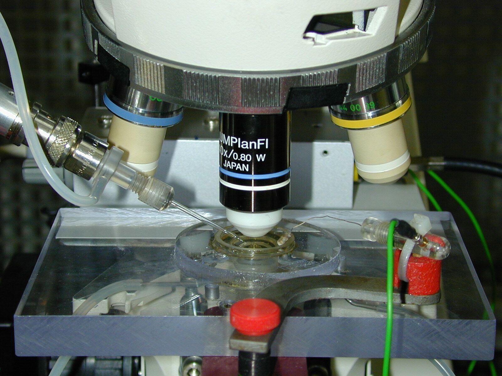 Set-up for carrying out patch-clamp recordings of cells.
© NMI
Set-up for carrying out patch-clamp recordings of cells.
© NMI
A breakthrough in the study of ion channels was achieved with the work of Erwin Neher and Bert Sakman in the 1970s for which they received the Nobel Prize for Physiology or Medicine in 1991. With the patch-clamp technique, Sakman and Neher presented a measurement method that enabled the flow of ions to be recorded (in the picoampere range) in individual ion channels. Manual as well as automated patch clamping requires a high level of skill with the device and the material under examination.
Today, many applications are based on the patch-clamp technique, for example in medicine. The major focus of research is on improving the diagnosis of diseases of the nervous system and muscles. There are genetic ion channel disorders such as the QT or the Brugada syndrome, in which the modification of proteins that regulate the transport of ions across the cell membrane results in the increased or decreased transport of potassium or sodium. The resulting change in the electrical properties of the heart muscle can lead to dangerous arrhythmias, ventricular fibrillation and cardiac arrest in people with otherwise completely healthy hearts. Here, the patch-clamp technique is used for the detailed analysis of the modified channels.
Companies and institutions are extremely interested in the further development of the patch-clamp technique. For example, some of these companies and institutions have optimised automated patch-clamp techniques to increase drug efficiency. Automated planar patch-clamping involves the application of a cell suspension on a chip with several openings of at least four micrometres in size. This arrangement enables several data points to be measured simultaneously, which is of huge value for the high-throughput drug discovery approaches used by the pharmaceutical industry.
Multielectrode arrays fuel hope
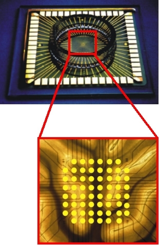 Multielectrode array with cell culture dish. The section shows the electrode array
© Rühe group
Multielectrode array with cell culture dish. The section shows the electrode array
© Rühe group
Meanwhile, neural interfaces that connect neurons with electronic circuits known as multielectrode arrays (MEAs) have become available. There are implantable MEAs which are used in vivo (in the living system) and non-implantable MEAs for use in in-vitro research involving cell cultures. While the benefits of in-vivo arrays are their high spatial resolution and the recording of individual nerve cell signals, the use of in-vitro arrays is non-invasive as their application does not result in damage to the cell membrane.
In-vivo MEAs are particularly used as cardiac and brain pacemakers; they are also used as cochlear implants, which are hearing prostheses for deaf people whose auditory nerve is still intact. As MEAs provide insights into neural processes such as perception and memory formation, they can be helpful in the diagnosis of diseases such as epilepsy and depression. A particular focus is also on the treatment of amyotrophic lateral sclerosis (ALS). However, only time will tell whether the restoration of motor control can be achieved with multielectrode arrays.
Stephanie Heyl - 01.07.2013
© BIOPRO Baden-Württemberg