Artificial intelligence in ophthalmology
Retinal diseases such as age-related macular degeneration (AMD) are now treatable. However, it is hard to predict individual disease progression. A group of researchers at the University Eye Centre in Freiburg are currently developing a new system which is hoped will allay fears and improve therapy planning. The system uses artificial intelligence to predict therapeutic outcome from image and patient data. Initial results are already available.
Retinal diseases, especially wet age-related macular degeneration (AMD), are the main cause of blindness and impaired vision in the developed world. Around six million people suffer from AMD in Germany alone. AMD develops when a misguided process destroys the retina, starting from the yellow spot (macula) in the centre of the visual field. Left untreated, the disease can very quickly lead to blindness. The ability to read, drive a vehicle or recognise faces is then so severely limited that those affected find it very hard to live normal lives.
Vascular endothelial growth factor (VEGF) therapy is an effective treatment option for AMD that has been used by ophthalmologists for about ten years now. The patient receives several injections of the medication – the VEGF inhibitor – into the vitreous body at agreed intervals. The progression of the disease can be slowed down and, in the best case scenario, even halted.
From deep learning to therapy predictions
 Prof. Dr. Andreas Stahl and his Angiogenesis research group at the University of Freiburg's Eye Centre are developing a system to predict treatment success of retinal diseases.
© private
Prof. Dr. Andreas Stahl and his Angiogenesis research group at the University of Freiburg's Eye Centre are developing a system to predict treatment success of retinal diseases.
© private
"Injection into the eye is an unpleasant procedure for the patient, which is required once a month in the initial stages of treatment," explains Prof. Dr. Andreas Stahl, managing senior physician and head of the Angiogenesis research group at the University Eye Centre in Freiburg. The clinic has years of experience with this type of AMD treatment, but some aspects of the disease still remain elusive. "Unfortunately, due to the very heterogeneous nature of the disease, we have not yet come up with an effective way to find out how often patients need to be injected and how their visual acuity will develop. We have had patients who needed only three treatments, while others have required over 60 treatments. This lack of predictability is unsatisfactory for doctor and patient alike."
The physicians therefore started thinking about developing a system that could predict how many injections would be needed and how successful they would be. This resulted in the BMBF-funded collaborative project "Therapy prediction through OCT and patients’ demographic data in the field of ophthalmology (TOPOs)", in which the University Eye Centre has been working for nearly two years with computer scientists from the University of Rostock and the University of Mittweida and the Freiburg text mining specialist Averbis GmbH. The researchers use deep learning to screen patients' medical records and image findings for information that allows personalised predictions to be made about individual disease progression in AMD and related conditions such as diabetic macular edema or retinal venous occlusion.
A wealth of information in OCT image data
Important starting materials are images taken using optical coherence tomography (OCT). "OCT gives us high-resolution images of the retina, non-invasively and without harmful radiation. Together with the VEGF inhibitors, this procedure has revolutionized retinal therapy in recent years," said Stahl. "And because the images harbour so many details that a human brain cannot fully comprehend, we wondered if the image data might contain subtle information that could tell us something about the aggressiveness and progression of the disease."
The OCT images are evaluated together with fully anonymised clinical information from medical reports. "We do not yet know if additional information - such as whether the patient has suffered a stroke or heart attack - could be relevant," says Stahl. "Maybe this is a pattern that correlates with the course of treatment, but we have so far been unable to identify a pattern." Averbis GmbH is tasked with analysing clinical texts by filtering out relevant information from large amounts of data. The medical reports do not have to be standardised, but are automatically analysed and evaluated in context by intelligent search systems.
Neural networks training using learning sets
The image data can now be analysed automatically thanks to a machine learning method known as deep learning. This part of the project is undertaken by the Department of Media Informatics at the Mittweida University of Applied Sciences (led by Prof. Dr. Marc Ritter). Artificial neural networks scan each image for possible early indicators of the further course of the disease. The neural networks are able to detect hidden relationships between visual findings and the future course of vision, provided that they are trained with a large amount of suitable visual material. By repeating this with the very heterogeneous disease trajectories of many thousands of patients, the algorithm perfects itself until it can establish a combination of initial findings and probable result, even for unknown data sets.
As a result, large structured data tables are created which may conceal important relationships related to the progression of the disease. To make this information visible, researchers from the Department of Computer Graphics (led by Prof. Dr. Paul Rosenthal) at Rostock University have developed an interactive visual software tool based on an individual patient's history, for example, whether an acute loss of visual acuity is actually related to the progression of retinal disease or instead is caused by a concomitant other eye disease, such as a cataract - information that is essential for further prognosis.
Machines will not replace humans
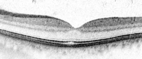 High-resolution images of the retinal layers have revolutionised the diagnosis and treatment of retinal diseases in recent years. Here: OCT image of a normal finding.
© Prof. Stahl
High-resolution images of the retinal layers have revolutionised the diagnosis and treatment of retinal diseases in recent years. Here: OCT image of a normal finding.
© Prof. Stahl
The researchers are keen to emphasise that their approach is not an automated therapy, but a therapy support: "If a patient knows that there is a chance of halting the progression of the disease after just three injections, treatment is no longer so scary and we can therefore encourage the patient to continue the unpleasant therapy,” Stahl points out. "Conversely, we can better prepare patients with less favorable initial findings for a potentially longer course of treatment. Knowing which way to go can significantly improve patient compliance. Artificial intelligence can help the ophthalmologist plan therapy, but does not replace it.”
The initial results give the project partners good reason to believe that they are on the right track. "We are currently at a point where we are already getting pretty good estimates of actual values and what may be early indicators from the system," says Stahl. "In the third year of the project, we hope to build on current results and come up with valid values for long-term prognoses. This is a big challenge, but we remain optimistic.”