The NanoLYRIC project: nanoparticles as drug carriers
Radiation-activated local chemotherapy
The reason why conventional chemotherapy is associated with severe side effects is because cytotoxic drugs act on the whole body. Researchers at the Karlsruhe Institute of Technology (KIT) and the University Medical Centre Göttingen, in collaboration with Aalen University of Applied Sciences, are developing a novel approach in which a chemotherapeutic agent encapsulated in nanoparticles is released locally and selectively only upon targeted irradiation of the tumour.
Cytostatic drugs are highly effective agents that inhibit or slow cell proliferation. They are primarily used in cancer therapy to suppress tumour cell growth or induce cell death. However, because these drugs are administered systemically – either by intravenous infusion or orally – rather than selectively targeting cancer cells, they also affect healthy cell proliferation throughout the body. As a result, treatment often leads to significant side effects. Tissues with a high cell turnover rate, such as hair follicles, bone marrow and the mucous membranes, are particularly susceptible to these toxic effects.
"We aim to increase the specificity of chemotherapy and thereby reduce adverse effects on healthy tissue," says Prof. Dr. Andreas Walter from the Centre for Optical Technologies (ZOT) at Aalen University of Applied Sciences (HSAA), describing the objectives of the NanoLYRIC collaborative project (short for: Nanoparticle-Based Local Hybrid Radiotherapy and Radiation-Activated Chemotherapy). Researchers from HSAA, the Karlsruhe Institute of Technology (KIT) and Göttingen University Medical Centre (UMG) are developing and validating a novel therapeutic approach based on gold nanoparticles (NPs) loaded with a chemotherapeutic agent. The drug is released locally only after targeted irradiation. "By combining chemotherapy with X-ray-based radiotherapy, we aim to increase treatment specificity and improve therapeutic efficacy," Walter explains.
Interdisciplinary research enables characterisation across different scales
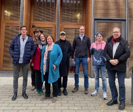 The partners in the NanoLYRIC project in front of the Centre for Optical Technologies building in Aalen, from left to right: Prof. Dr Andreas Walter, Prof. Dr. Christian Dullin, PhD student Louisa Herbsleb, Prof. Dr. Frauke Alves, PhD student Paulina Chojnacka, Prof. Dr. Christian Neusüß, PhD student Constantin Bauer, Prof. Dr. Claus Feldmann. © Andreas Walter
The project has been funded since early 2025 by the Carl Zeiss Foundation through its CZS Transfer programme. Prof. Dr. Claus Feldmann from the Institute of Inorganic Chemistry at the Karlsruhe Institute of Technology (KIT) is developing and synthesising the gold nanoparticles (NPs). At Aalen University of Applied Sciences (HSAA), Prof. Dr. Andreas Walter is using high-resolution microscopy techniques to investigate the uptake and intracellular localisation of the nanoparticles. His colleague, Prof. Dr. Christian Neusüß from the Department of Analytical Chemistry, is using capillary electrophoresis–mass spectrometry (CE–MS) to quantify the chemotherapeutic agent released after irradiation, as well as its intracellular metabolites. The therapeutic efficacy – and thus the approach’s potential for treating tumours – will ultimately be evaluated at the Göttingen University Medical Centre (UMG) by Prof. Dr. Christian Dullin (Experimental Imaging), Prof. Dr. Frauke Alves (Translational Molecular Imaging) and Prof. Dr. Stefan Rieken (Department of Radiotherapy and Radiooncology). The team will assess the treatment in breast cancer cell cultures, computational simulations and preclinical mouse models.
The researchers met through the interdisciplinary EU-funded research project COMULIS, which promotes correlated multimodal imaging and analysis across multiple spatial scales. By adopting a correlative approach – that is, combining complementary imaging techniques – the project aims to overcome the limitations of individual methods and enable comprehensive characterisation of nanoparticle-based therapies across several orders of magnitude. As Prof. Dr. Andreas Walter explains: "Our goal is cross-scale imaging – from tissue to individual cells and ultimately to the molecular level. The NanoLYRIC project aims to develop and implement this concept using novel nanoparticles as a specific application example."
Novel inorganic-organic hybrid nanoparticles
The innovative therapeutic approach developed by Feldmann and Dullin is based on the inorganic–organic hybrid nanoparticles (NPs) created by the Feldmann research group.1) These nanoparticles have a salt-like structure, consisting of inorganic cations and negatively charged molecules of an organic chemotherapeutic agent. Building on this concept, the current project is developing novel gold nanoparticles loaded with chemotherapeutic agents. During radiotherapy, the nanoparticles absorb increased amounts of X-ray radiation within the tumour, triggering the drug’s local release. This strategy is designed to enhance the efficacy of cancer treatment and minimise damage to healthy tissue.
In principle, the researchers assume that, even after systemic administration, the nanoparticles (NPs) will tend to accumulate in tumour tissue due to the enhanced permeability and retention (EPR) effect. Blood vessels in tumours are more permeable than in healthy tissue and clearance of foreign substances is impaired, which promotes the retention of nanoparticles.
High-resolution CLEM technology
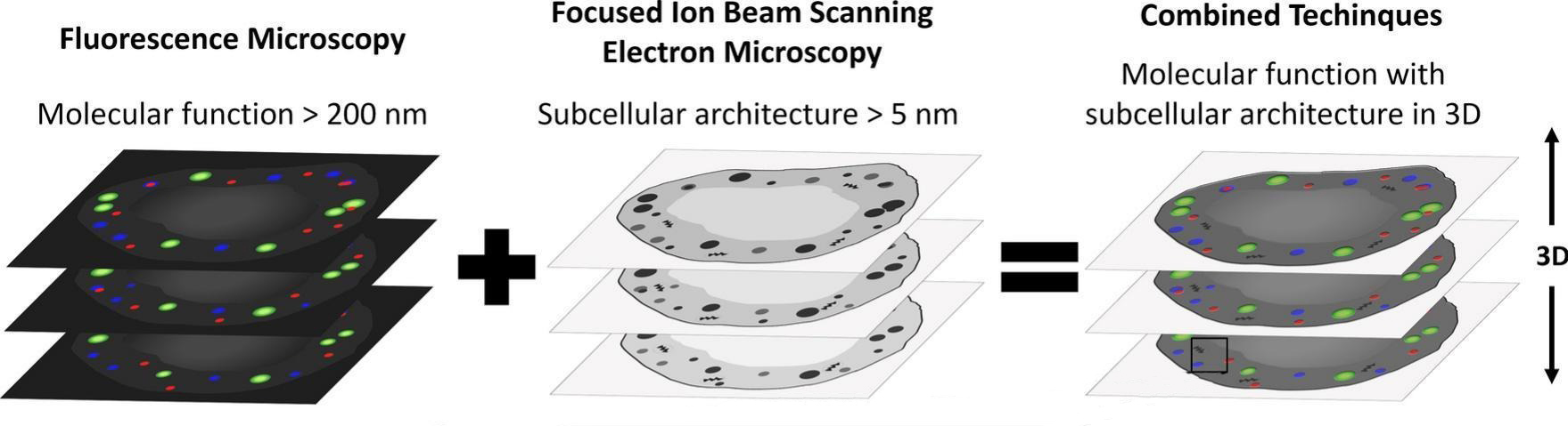 Correlative light and electron microscopy (CLEM) combines high-resolution three-dimensional (3D) fluorescence microscopy with 3D electron microscopy. This integrated approach enables researchers to visualise nanoparticles within their subcellular environment and to trace their intracellular journey from uptake and trafficking to their eventual degradation. © "Graphical Abstract“, Herbsleb et al., Nanomedicine, https://doi.org/10.1016/j.nano.2025.102872, CC-BY 4.0 (https://creativecommons.org/licenses/by/4.0/), modified by R. Menßen-Franz
After just over a year, the key methods for characterising the nanoparticles (NPs) have largely been established, according to Prof. Dr. Andreas Walter, the project coordinator. His team will be further developing the optical imaging techniques and is applying the innovative correlative light and electron microscopy (CLEM) approach, a combination of fluorescence microscopy (FM) and electron microscopy (EM). While FM can be used to visualise fluorescence-labelled nanoparticles, EM provides high-resolution three-dimensional images of cell and tissue ultrastructure. By integrating both techniques, researchers can directly visualise specific molecules and nanoparticles within their subcellular environment and track their intracellular path from uptake and trafficking through to eventual degradation.
However, because the nanoparticles (NPs) are only around 60 nm in diameter, the resolution of the fluorescence microscope must exceed the optical microscopy’s standard diffraction limit of approximately 200 nm. "We build our microscopes ourselves and have now established a high-resolution 3D-CLEM workflow," says the physicist. In a paper published in the journal Nanomedicine in October 2025, the researchers demonstrated in detail the cellular uptake, intracellular distribution and degradation of the particles using zirconium nanoparticles as a model system.2)
This therapeutic approach has many advantages
Both the composition of the hybrid nanoparticles (NPs), which consist of gold plus a chemotherapeutic agent, and the combination of radiation-activated chemotherapy with radiotherapy are a novel concept that has never been used before and offers several advantages. Synthesising this patent-protected class of nanoparticles is straightforward and can incorporate a wide range of cytostatic drugs as well as other therapeutic agents such as antibiotics. Owing to their salt-like structure, the nanoparticles also achieve an exceptionally high drug loading of 70–90 wt%.
The irradiation required to release the active substance can be carried out using equipment routinely available in hospitals. Furthermore, rather than being limited to a specific type of tumour, this therapeutic approach is in principle suitable for all solid tumours. However, as the chemotherapeutic agent acts exclusively at a local level, it cannot reach metastases.
The interdisciplinary research team is confident that by the end of the funding period it will be able to demonstrate – based on preclinical data – that the combined chemotherapy–radiotherapy approach both enhances therapeutic efficacy and reduces treatment-related side effects. The researchers plan to build on these findings to advance the clinical translation of the technology through a subsequent funding programme. Walter emphasises: "This innovative project is founded on excellent basic research, whose full potential can only be realised through close interdisciplinary collaboration."
