Neurosciences
Mesh microelectrode arrays: research with brain organoids on a new level
How does the brain work? Brain organoids are derived from pluripotent stem cells and regarded as valuable model systems that can depict some aspects of neurological functioning. Dr. Peter Jones from the Institute of Natural and Medical Sciences at the University of Tübingen, together with Dr. Thomas Rauen from the Max Planck Institute for Molecular Biomedicine in Münster, has taken organoid research to a new level. His novel mesh microelectrode array (Mesh-MEA) greatly improves the growth and electrophysiological analysis of tissue.
Our understanding of neuropsychiatric diseases such as schizophrenia and Alzheimer's is far from complete and the search for therapies is laborious, as research into the human brain is only possible to a limited extent. Clinical studies provide insights into brain waves but no information about metabolic processes. Representative animal experiments are often inaccurate, as humans and animals differ in many ways. Alzheimer's and Parkinson's disease are difficult to modulate in animals, as they do not occur naturally in mice. In addition, scientists are endeavouring to avoid or at least significantly reduce the use of animals for research purposes. As an alternative, brain organoids offer promising prospects for science and the Max Planck Society is heavily promoting organoids as alternatives to animal testing in neuroscience as part of the “White paper – animal research in the Max Planck Society” project.
Self-organising 3D tissues
 Dr. Peter Jones is developing a mesh microelectrode array (mesh-MEA) at the NMI Natural and Medical Sciences Institute at the University of Tübingen, which greatly improves the investigation of brain organoids. © Dr. Peter Jones, private
An organoid is similar to an organ and bridges the gap between classical 2D cell cultures and in vivo vertebrate models. The organoid is physiologically more relevant than 2D cell cultures and more accessible for use in analyses than the brains of live mice. Organoids are produced by dedifferentiating cells from human skin biopsies into pluripotent stem cells in vitro. Special growth factors are then added to the culture medium in the Petri dish to enable the dedifferentiated cells to develop into nerve or glial cells in a similar way as those in the brain. The cells differentiate into these specific tissues with a characteristic cellular architecture in a self-organising manner as they would in the body. 3D spheres no larger than a pea grow autonomously from the 2D cell cultures. This can imitate certain functional aspects of an organ. Findings show that the nerve cells in brain organoids communicate with each other and even develop circuits. While it is not possible to represent the entire brain in all its complexity, the consequences of certain genetic defects or substances on human brain tissue can be observed. This is particularly interesting as the organoid cells involved originate from biopsy material taken from patients and therefore reflect their individual genetic repertoire.
Contactless measurement of electrical cell activity
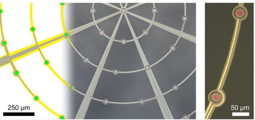 Left: The hanging spider web-like mesh with integrated microelectrodes (green) shows that the regions between the polymer filaments are open. Right: Two microelectrodes in a single filament.
Source: https://doi.org/10.1101/2020.09.02.279125; Creative Commons Attribution 4.0 International License.One particular difficulty is capturing the electrophysiological information of spherical organoids using planar measurement technology, as the spherical organoids can only contact a few electrodes when placed on a planar MEA. Planar microelectrode arrays (MEA), which can only detect activity up to a distance of 100 micrometres from the MEA surface, are only suitable to a limited extent for structures like organoids that measure one to three millimetres in diameter. "We only capture the activity of one cell at the site where it accidentally happens to lie on the electrode," explains Dr. Peter Jones from the Natural and Medical Sciences Institute at the University of Tübingen (NMI). Previously, if you wanted to find out more about the internal electrical activity of the nerve cells in the organoid, you had to prick the tissue with a probe or cut it open. However, the results were arguable due to the resultant tissue damage.
Another problem relates to the propensity of cell cultures to stick to the substrate on which they grow and then fail to develop three-dimensional structures. A few years ago, Jones was asked by the Max Planck Institute (MPI) in Münster to develop a process that would work better for organoid research. The engineer, who has specific knowledge of micro- and nanosystems technology, had an idea that could easily work in practice. His team invented a microelectrode construct made of polyimide that contains 61 microelectrodes and hangs freely in space like a spider's web. The construct looks like a tiny hammock that hangs high above the bottom of the Petri dish. "What is exciting is that the organoid is dynamic, it arranges itself and grows unhindered through and around the hammock," says Jones. The mesh electrode array therefore becomes completely embedded in the organoid. This allows action potentials (as a sign of electrophysiological activity) to be recorded extracellularly and without contact. At the same time, long-term recordings can be made at many points inside the organoid over the course of a year as long as the organoid continues to grow. "The activity of a cross-section of the organoid can be recorded without interfering with its development," says Jones. In addition to providing more information, organoids can more easily be supplied with nutrients and oxygen than 2D cultures, as these can be added from every angle. According to Jones, selecting the right processes was a challenge, as the materials had to be biocompatible and the conductive electrodes of the mesh-MEA had to remain electrically active in liquid.
Individual organoids open the door to personalised medicine
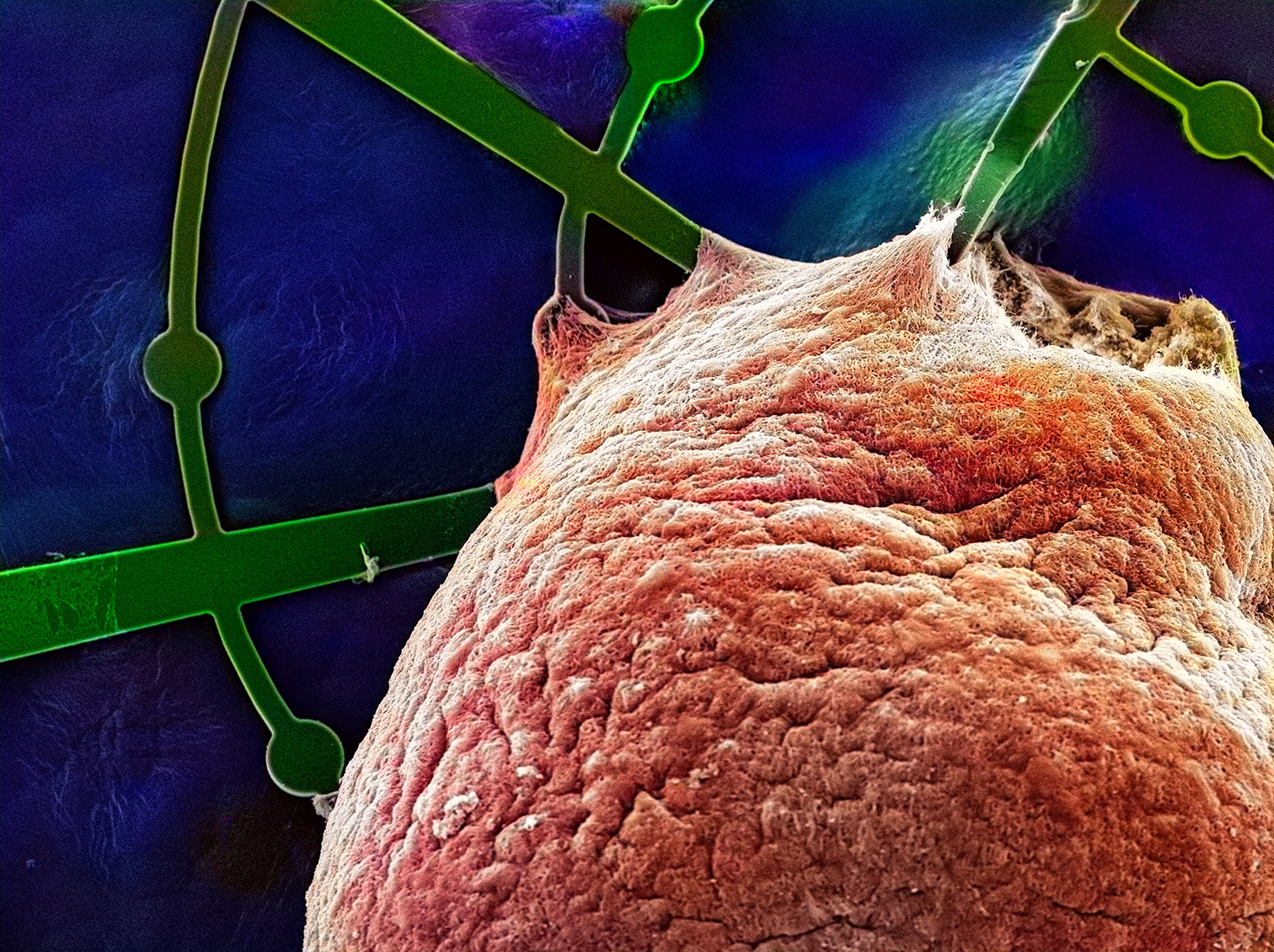 A brain organoid made of human pluripotent stem cells (shown in red) grows around and through the mesh microelectrode array (shown in green). The mesh filaments and the round microelectrodes are partially hidden in the organoid. © Max Planck Institute for Molecular Biomedicine/ Thomas Rauen, Katherina Psathaki
Research on the living brain is neither possible nor ethically justifiable. In fact, researchers rarely have the opportunity to analyse brain tissue. "With human brain organoids, we now have the opportunity to conduct in vitro research without any ethical issues," says the engineer. They allow the researchers to gain deeper insights into early brain development and the development of neurological diseases. They can be used to investigate the effects of toxic or growth-promoting substances, viruses and other germs on nerve cells, and follow their evolution. The researchers are primarily measuring a tissue’s overall activity, regardless of which cell is active and when. Depending on which substance is used, neurotoxins either cause tissue activity to decrease or increase, and can therefore be used as control substances.
Until now, there have been no physiological microcircuits in cell cultures. In the future, however, organoids might form such circuits. This has the potential to improve the models used. It may be possible, for example, to develop a "learning" model to represent effects such as long-term potentiation (LTP). In addition, it would also be conceivable to test drugs in different dosages in parallel experiments or to link different organoids with each other. One particular challenge is developing models for neuropsychiatric diseases such as schizophrenia and autism, which are correlated with developmental disorders of the nervous system. These very heterogeneous diseases require personalised approaches. Organoids produced from patient-specific tissue would provide individualised insights into disease pathology and specific drug action.
Huge medical potential
A tiny isolated brain organoid in a Petri dish is of course not the same as a brain that has grown inside the body for years. The density and complexity of living nervous tissue cannot be achieved in vitro. Although the organoid initially has a very similar microstructure to its neural counterpart, the longer the culture grows, the more it diverges from what actually happens in the brain. Further specialisation of the organ would require sensory input from the sensory organs and messenger substances that do not exist in the organoid. Subsequent processes, such as neurodegenerative diseases, take a very long time and cannot currently be represented. Brain organoids can therefore help to reduce the number of animal experiments in drug efficacy and toxicology studies but cannot currently eliminate them.
Jones' working group is also carrying out research into the development of microimplants in organs such as the pancreas, which would be used to record activity and potentially ensure long-term stable measurement of blood glucose levels. In the "nanodiag BW" future cluster, researchers are working on solid-state nanopores that can detect and measure individual biomolecules such as proteins or DNA. For Jones, neuroscientific research is necessary to understand the brain, as much is still unknown and there is still no treatment for some diseases. "If we want to make progress on neurodegenerative diseases, we need good tools," says the scientist.