Medical image analysis
Powerful AI systems using synthetic training data
AI systems for image analysis are only as good as the data on which they are trained. The Göppingen-based start-up MIRA Vision has developed a novel method for generating synthetic, photorealistic images, enabling the efficient creation of large, high-quality training datasets. An intuitive platform also allows researchers to evaluate microscopy images with ease.
Many medical diagnoses rely on imaging techniques such as MRI, CT scans, X‑rays and microscopy. These images contain a wealth of information, but interpreting them is often complex and time‑consuming. Computer programmes powered by artificial intelligence (AI) are increasingly being deployed to support clinicians. For these AI systems to work reliably, they must first be trained on a large number of ground-truth examples, which are produced by experts painstakingly marking relevant structures in a labor‑intensive process called annotation. "Collecting and annotating the necessary images is extremely time‑consuming and expensive," explains Leonid Mill, CTO of the start‑up MIRA Vision. "That is why we generate synthetic, photorealistic images using a novel process."
Analysis of microscopy images requires high-quality training data
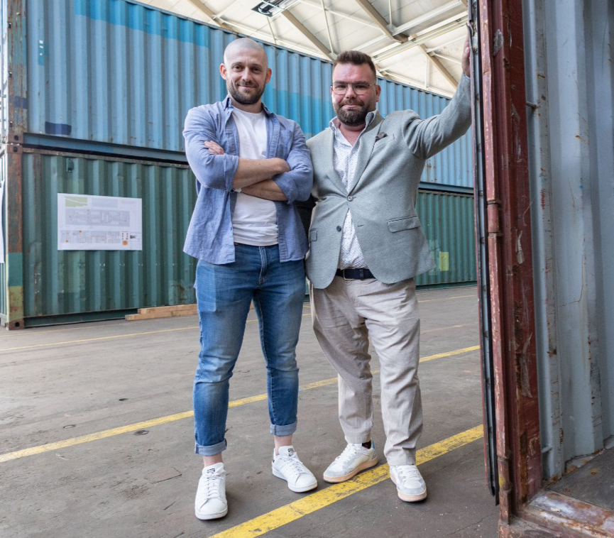 Leonid Mill (left) and Lukas Mürdter (right) from MIRA Vision use synthetic photorealistic images to train AI systems to analyse microscopy images. © NWZ
AI-based applications have great potential, particularly in histology – the microscopic examination of tissue samples. The algorithms segment microscopy images, assigning each pixel to an object. This enables tiniest structures and complex patterns to be detected, allowing pathological changes to be identified precisely, objectively and quickly.
However, the traditional path to a suitable AI system is arduous. Analysing detailed microscopy images requires a large amount of high‑quality training data. When pathologists manually annotate selected raw images, they label each individual structure with information – for example, tissue type, benign or malignant status, or staining intensity – a process that can take several days per image. To create a reliable algorithm for something like tumour detection, millions of such images or image patches are needed, ideally representing many variants of healthy and diseased tissue. "To make matters worse, there can be significant differences in colour and contrast between microscopes from different manufacturers," says Lukas Mürdter, CEO of the Göppingen‑based company.
"The AI must therefore be trained with data from each individual microscope."
Extensive datasets from synthetic images
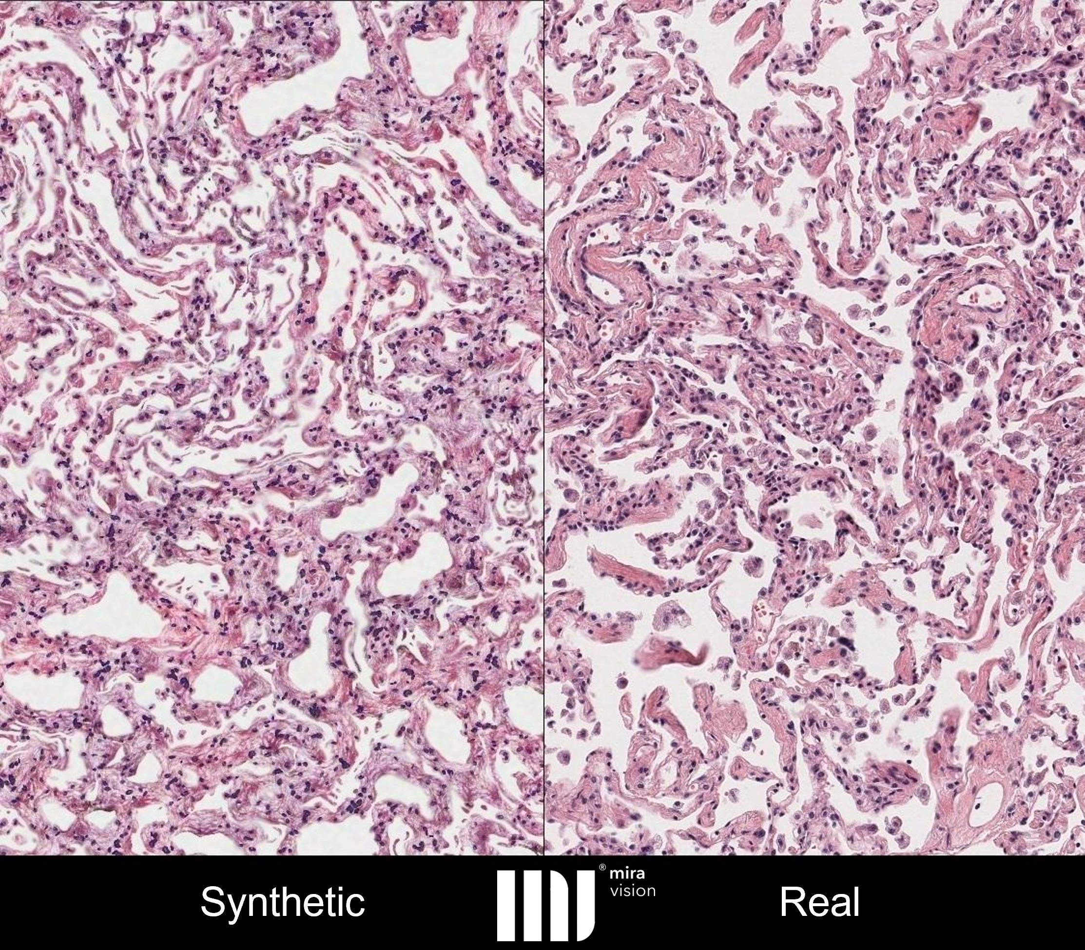 The synthetic image of stained lung tissue (left), generated using MIRA Vision’s innovative process, is indistinguishable from the real microscope image (right). © MIRA Vision Microscopy GmbH
Mill also faced these challenges during his time with Prof. Dr. Andreas Maier at the Department of Pattern Recognition at Friedrich‑Alexander University Erlangen‑Nuremberg (FAU), where he worked on several research projects involving AI‑supported image analysis in the medical field. The mechatronics engineer explains: "I developed an alternative technology to obtain the required data more quickly and with far less effort. Using this technology, we can now generate photorealistic – in other words, synthetic – images that are indistinguishable from real microscopy images."
At the start of the process, specialised software is used to manually simulate individual tissue components – such as cells, fibres, connective tissue, artefacts and colour variations – without using generative AI. Original images and illustrations from textbooks, scientific publications and the internet serve as templates. These basic building blocks are then combined to create images of stained tissue sections with varied textures. In an iterative process with experienced pathologists, the images are refined until even experts are unable to distinguish them from real microscopy images.
Countless datasets based on the simulations can be created in a very short time and used to train AI models efficiently. "The great advantage of our technology is that each pixel is defined by a mathematical function," Mill emphasizes. "This lets us generate every conceivable variation – the ideal basis for a reliable AI system." By avoiding real patient data, the complex issue of data protection is also eliminated, adds Mürdter.
Mill and his colleagues at FAU Erlangen–Nuremberg demonstrate the effectiveness of this novel approach in a publication that appeared in March 2025. 1) Using stained muscle preparations as an example, the researchers show that an AI system trained on synthetic images can even outperform one trained on real data. The segmentation – the precise assignment of individual image pixels to objects – is higher quality and achieves the same level of results as medical professionals.
User-friendly MIRA AI platform
The innovative process produced its first promising results in late 2020. When Mill told his brother‑in‑law Mürdter – a trained business economist, then managing director of an IT service provider – about it, he saw the technology’s business potential, and the two founded MIRA Vision Microscopy GmbH in May 2021. With financial support from private investors and public funding – for example through the European BioMan4R2 project and a HighTech BW innovation voucher – the web‑based MIRA AI platform was developed over subsequent years. It makes automated image analysis accessible to many users, currently in the field of muscle histology. Researchers can upload their own images and have them quantified without any particular prior knowledge. Mill explains: "The system accurately records how many cells and cell nuclei are present and the proportion of connective tissue. All these metrics provide important information about what is happening in the tissue." The information can be used to determine the effects of drugs, diseases, or other factors on tissue. Various university research groups in Germany, Austria and Canada have already successfully applied the platform.
Research institutions such as the German Aerospace Center (DLR) have also benefited from the technology. MIRA Vision analyzed all muscle-tissue sections for the AGBRESA bed-rest study conducted by NASA, ESA and DLR, which investigated the effects of artificial gravity on astronaut health.2)
Wide range of applications
The patented method for training AI systems is globally unique. In 2023, the company, which now has ten permanent employees, won the Göppingen Innovation and Entrepreneurship Award, and in 2024 it was named AI Champion of Baden‑Württemberg. MIRA Vision is part of the renowned AI research consortium Cyber Valley and is a portfolio company of the Federal Agency for Breakthrough Innovations (SPRIND).
The start-up is currently working on projects in lung, breast and cervical histology. A detailed description of individual cells offers real added value to pathologists and can help identify disease markers. In the long term, the AI systems are intended for use in clinical diagnostics. In addition to quantification, the platform can classify cells based on morphological changes – an important step in disease detection. The ratio of healthy to diseased cells could also serve as a marker for disease progression.
The Göppingen‑based technology can be used to examine tissue samples and blood smears, for example to detect malaria‑infected cells. "There are only a few experts in malaria detection worldwide. We want to help ensure that everyone has access to affordable diagnostics," says Mürdter. "Collecting real data from around the world to train an AI would take years." Thanks to its innovative technology and broad network of experts, MIRA Vision can develop a powerful AI system for any area of application within a few weeks.