Imaging methods in medical diagnostics
Many different imaging methods are available these days and are used in almost all medical disciplines to visualise disease-related changes. Depending on the problem and the clinical picture, very different structural and functional parameters can be visually recorded for diagnosis and used for therapy.
Diagnostic imaging procedures are indispensable in modern medicine. The most widely known procedures – ultrasound, endoscopy, X-ray diagnostics, CT (computed tomography) and MRI (magnetic resonance imaging) – are each used many millions of times a year in Germany. These methods have been established for decades1), but new and further developments that improve diagnostic assessments and open up new areas of application are constantly being described. In addition, there are lesser known and even completely new imaging and hybrid techniques that combine the advantages of different methods. The following table endeavours to provide an overview of the key imaging methods currently available in medical diagnostics and their areas of application.
A number of research projects are being carried out in Baden-Württemberg that could determine the development of imaging methods in the future. For example, in the QSens Future Cluster, quantum sensors are being tested for their use in biomedical diagnostics. These sensors are orders of magnitude more sensitive than is possible in imaging today.2)3) Cluster partners in this project include the Universities of Stuttgart and Ulm as well as companies such as Boehringer Ingelheim Pharma, Rentschler Biopharma and NVision Imaging Technologies, a start-up in the field of quantum technology in which researchers from Ulm are involved.4) Using a different approach to quantum sensor technology, a sub-project of the Fraunhofer-Gesellschaft's lead project QMag is developing a method in which polarisers made of nanodiamonds are combined with biomarker molecules and used for MRI diagnostics. With the help of this quantum magnetometry, imaging could become ten thousand times more sensitive than in current MRI examinations.5)
Magnetic resonance imaging (MRI) and magnetic resonance angiography (MRA)
MRI is based on measuring the nuclear spin signals of protons of water in the magnetic field, and one of its advantages is that it does not require harmful radiation.6)7) Its disadvantages – greater inaccuracy, relatively speaking, and the long duration of the images – can be largely compensated for by new developments, meaning that MRI and magnetic resonance angiography (MRA), which visualises blood and lymph vessels, are now important diagnostic tools for a wide range of diseases.
Further developments of the FLASH (fast low-angle shot) technology most commonly used nowadays reduce the time required for MRI images to well below a second, making even film recordings of the beating heart or the blood flow in the brain possible.8) Sharper images are obtained by hyperpolarising the nuclear spin signals in strong magnetic fields9) or by combining MRI with optical microscopy.10) Multiparametric MRI (mpMRI) produces high-resolution cross-sectional MRI images as well as determining and evaluating other measured variables such as the movement (diffusion) of the water molecules and the blood supply (perfusion) with the help of intelligent image analysis methods. mpMRI has since become an important decision-making basis for therapy planning in diseases such as prostate carcinoma and breast cancer.11)12)
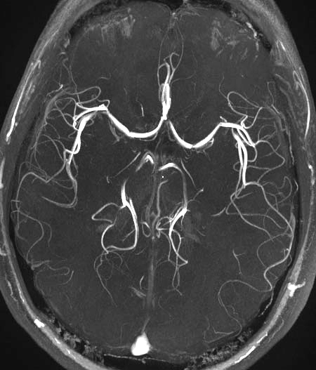 Visualisation of the cerebral vessels and the circulus arteriosus cerebri (circle of Willis) using time-of-flight magnetic resonance angiography (TOF-MRA) in a 4Tesla magnetic field. © SBarnes 12 Feb 2009; creative Commons BY-SA 3.0. Wikimedia Commons File Mra-mip.jpg.
Contrast agents are often used in MRI – especially MRA. In this way, even the finest ramifications of the vessels or the flow of fluid can be made visible by means of perfusion imaging.13) However, with phase-contrast MRA, different velocities of blood flow can be measured even without contrast-enhancing agents, rendering constrictions, bulges and blockages in arterial vessels visible. Time-of-flight MRA (TOF-MRA), which is used notably in examinations of the cerebral vessels, is based on the principle that blood freshly flowing into the vessel has a higher magnetisation than the surrounding tissue and is therefore imaged with a stronger signal. A new method for diagnosing brain tumours, which are characterised by a high glucose metabolism, tested by researchers from Tübingen and Heidelberg, also requires no contrast agent. Here, the changes in the magnetic field are measured during the proton exchange between glucose and water molecules.14)15)
While these methods rely on devices with high magnetic field strengths, which are only available in a few places16), intensive work is being undertaken on the development of miniaturised, compact high-performance MRI devices that are affordable for all clinics and diagnostic centres.17) In this context, the magnetic particle imaging method, which is still in an early trial stage, is interesting. Here, magnetic nanoparticles are applied to the organ structures under investigation and recorded by MRI.18)
Positron emission tomography (PET) as well as PET/CT and PET/MRI
PET has proven itself in cancer diagnostics because – unlike the established CT, MRI or ultrasound procedures – it can provide information about the metabolic activities of a tumour. In this way, metastases and tumour regressions (remissions) can also be detected after therapy.19) In the most modern devices, PET is combined with CT or MRI to form a hybrid procedure with higher image resolution, so that tumour changes can be detected earlier.20)-23) It is even possible to image individual leukaemia cells in patients’ bone marrow.24)
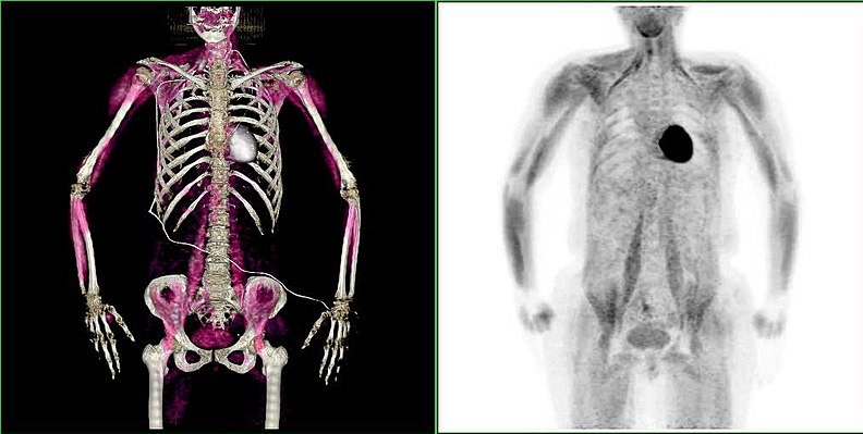 Positron emission tomography in combination with computer tomography (PET/CT). Image of the heart muscle after absorption of (18F)fluorodeoxyglucose under physiological conditions. © Hg6996 (Public Domain) https://commons.wikimedia.org/w/index.php?curid=9697305
PET uses radioactive tracer molecules that emit positrons in a beta+--decay, which, along with their antiparticles, electrons, immediately annihilate on the spot. The resulting pairs of gamma photons, which are emitted in opposite directions, are measured. This can then be used to calculate a picture of the exact location of the beta decay. The most commonly used tracer molecule is glucose, which has been labelled with a radioactive fluorine atom (18F). Like normal glucose, (18F)fluorodeoxyglucose is transported in the bloodstream and taken up by the cells, but is not completely metabolised, so that the fluorinated metabolic product accumulates in the tissue. Since many tumours metabolise glucose particularly intensively, they are clearly visible in the PET examination.25)26)
However, work is also underway to develop new radiodiagnostic compounds that, in combination with MRI, can detect tumour cells, bacteria, fungal cells and even individual cell structures. These will be used to plan and guide cancer immunotherapies.27)28) PET diagnostics using radiolabelled immune cells directed against cancer cells is still in the early stages of development.29)
About ten years ago, a PET diagnostic agent with a tracer molecule made of prostate-specific membrane antigen (PSMA) and radioactive gallium was synthesised for the precise detection of prostate cancer; it has now been further developed for cancer therapy by nuclear medicine researchers based in Heidelberg. For this purpose, the PSMA was coupled with an alpha or beta emitter, which destroys the tumour cells when the molecule binds.32) Such a combination of diagnostics and therapy in so-called theranostics is considered forward-looking; it forms one of the research priorities in the new Research Centre for Imaging and Radiooncology, which opened in Heidelberg in 2019.33)
However, PET has potential far beyond cancer diagnostics, and it is hoped that in future it can also be used for Alzheimer's and Parkinson's diseases as well as cardiovascular diseases.30)31)
Computed tomography (CT) and single photon emission computed tomography (SPECT)
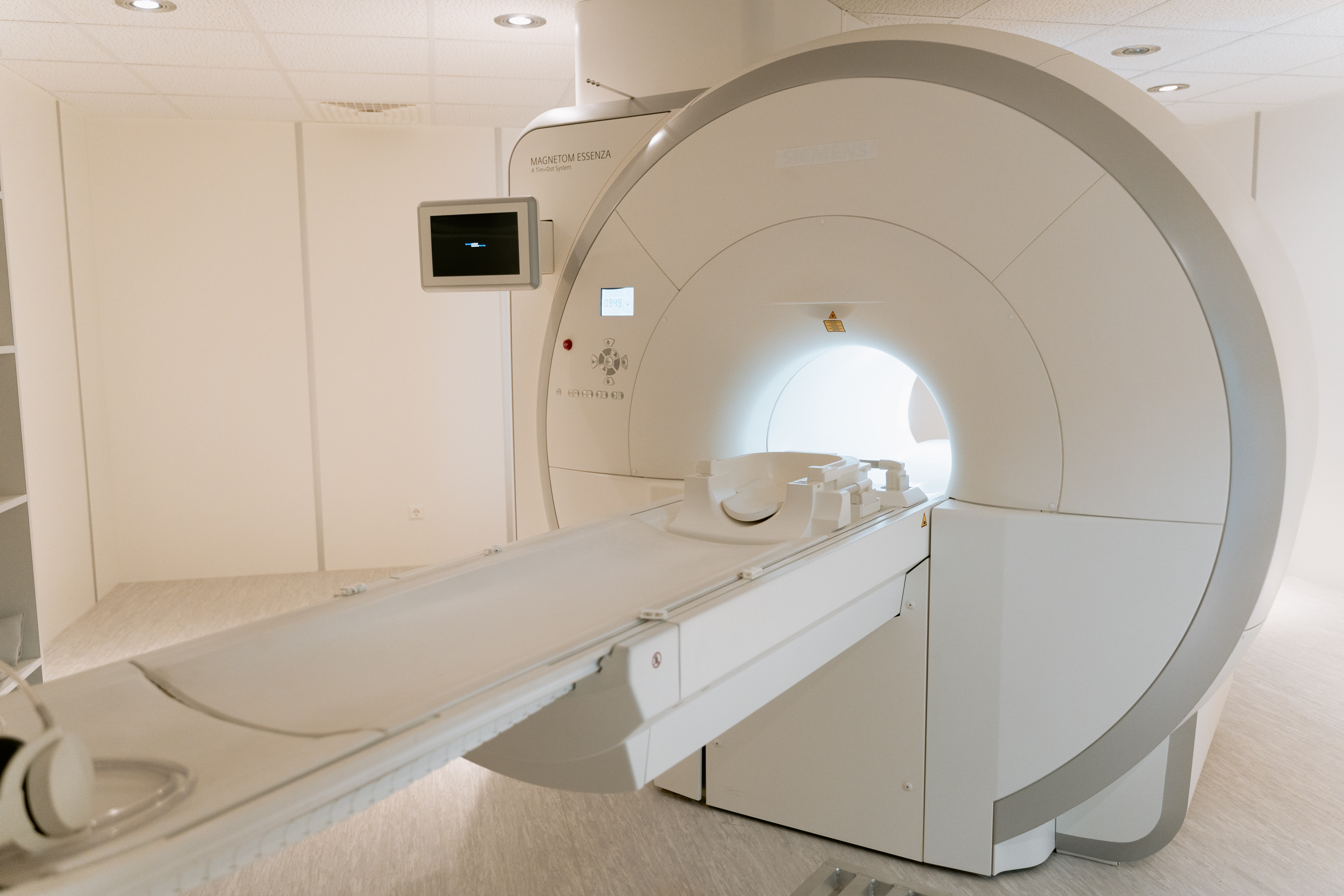 With a new type of detector system, images with higher spatial resolution can be genera-ted using computer tomography. © MART PRODUCTION / pexels
CT is perhaps the most important method for cancer diagnostics.34) With a new photon-counting detector system, images with higher spatial resolution can be generated so that, for example, bone metastases can be better detected.35) As part of the Forum Health Region Baden-Württemberg, this new technology is being tested for future therapy approaches in the Photon-Counting (PC)-CT Consortium (PC3) joint project at the University Hospitals of Freiburg, Tübingen and Mannheim. Other project partners in the consortium are Siemens and BIOPRO Baden-Württemberg. It is funded by the Baden- Württemberg Ministry of Economic Affairs.36)37)
SPECT, like PET, primarily depicts metabolic activities and is usually combined with CT in order to produce good images of structures. A weak gamma emitter with a short half-life is used as a tracer, mainly a technetium isotope. SPECT is now a standard procedure for measuring cardiac perfusion and myocardial function; it is cheaper and less complex than PET. However, SPECT is also widely used to diagnose brain diseases and to detect cancer metastases that differ in metabolism from healthy tissue.38)
Artificial intelligence in imaging
In CT, as in the other imaging procedures mentioned, one of the main problems is the processing and interpretation of the huge quantities of computer-generated images, which is almost impossible to perform manually and is very error-prone.39) Computer programmes for semi-automatic image segmentation are already being used. In a nationwide programme coordinated by the Freiburg University Medical Centre, work is being done on fully automated high-throughput image recognition using artificial intelligence (AI).40) A strategic initiative for image analysis and machine learning called the Joint Imaging Platform has also been established at all sites of the German Cancer Consortium DKTK.41) Advances in digital imaging have not only enabled the development of mobile devices for radiological practice,42) but also the display of complex 3D models of blood vessels, which are needed for navigating catheters in the brain.43) By merging IT, microrobotics and imaging, it has been possible to introduce microrobots into the smallest capillaries and make them visible from the outside; it is hoped that they can be used as theranostic instruments in the future.44)
Contrast agents and molecular imaging
As regards MRI, CT and X-ray diagnostics, the informative value of the images can often be improved by administering contrast agents. They are also widely used in ultrasound examinations, which are probably the most common imaging procedure in medical practice. The development of new contrast agents for special applications, which is associated with strict approval procedures, is a separate, indispensable sector of modern diagnostics.45)46) For example, by using highly specific molecular markers in the contrast agents, cancer metastases can be visualised on the basis of the bound marker molecules – an example of dynamically developing molecular imaging methods.47)48)
Endoscopy
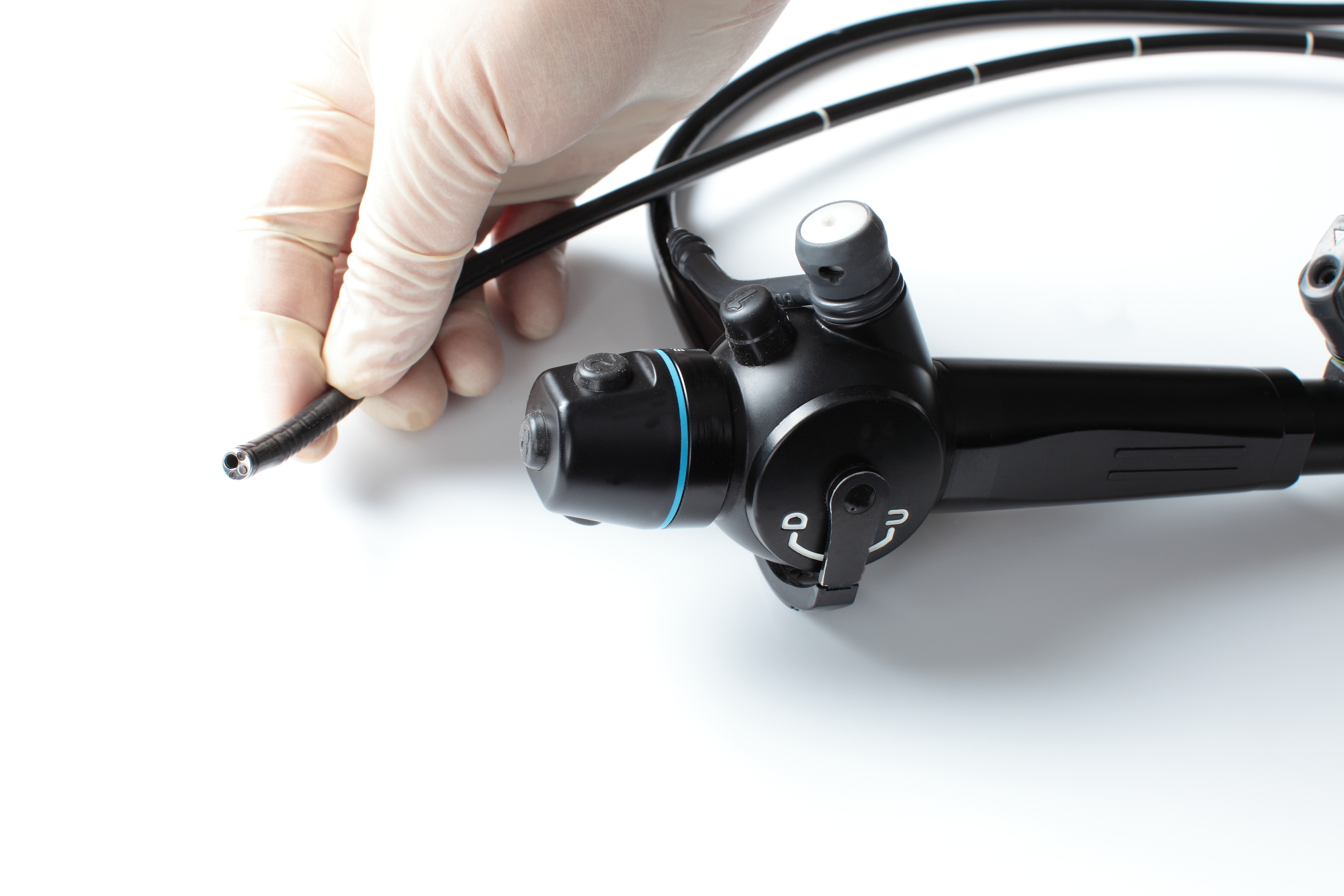 Endoscopes can be used to examine internal cavities in the body with optical probes. © nobasuke / Adobe Stock
New developments in endoscopy, which can be used to examine internal cavities in the body with optical probes, are heading in two main directions. The first involves devices with micro-optics made of fine glass fibres are increasingly being miniaturised, so that today even the inner walls of the smallest vessels can be scanned for plaque deposits with a laser beam. The examination is not done directly; instead it uses a digital micro-camera and computer-assisted image processing.49) Capsule endoscopy, which is being developed in a joint project coordinated by Ovesco Endoscopy AG, works in a similar way. This is an externally controllable capsule equipped with a camera, battery and transmitter that is swallowed by the patient undergoing diagnostic gastroscopy.50) With the camera images of normal endoscopes, tumours and healthy tissue can often barely be distinguished. The Mannheim-based start-up Thericon is therefore developing a multispectral imaging technology in which the images of different spectra are superimposed for better detection.51)
The second major development trend in endoscopy is to combine diagnostic optics with a working channel for flexible tools to take biopsy samples for laboratory diagnostics, inject active substances and destroy or remove diseased tissue. Two well-known applications are exemplary: firstly, an endoscopy of the ciliary body in the eye with simultaneous sclerotherapy with a laser beam to treat glaucoma. Secondly, a colonoscopy (examination of the large intestine) with simultaneous removal of polyps of the intestinal mucosa that might be early tumour stages.52) Thus, preventive colonoscopy from the age of 55 onwards, as one of the most successful measures of cancer control ever, has significantly reduced the number of deaths from intestinal cancer.